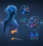




Weight resistance in menopause is the result not of the calories you take in but of the balance among estrogen, insulin, and the gut microbiome.
Summary
Weight resistance in menopause often stems not from a calorie surplus but from the metabolic shift set off by the drop in estrogen. The loss of estrogen affects insulin sensitivity, the gut microbiome, and muscle tissue all at once. That is why a sustainable approach rests not on a single piece of advice but on a framework that assesses these systems together.
In this article
- Why does fat distribution change when estrogen falls in menopause?
- How does insulin resistance begin in menopause, and how is it recognized?
- How do the gut microbiome and the estrobolome affect weight in menopause?
- What other factors influence weight resistance in menopause?
- How should nutrition be arranged in menopause?
- What does weight in menopause really tell us?
Not being able to lose weight in menopause is one of the most common complaints women raise. Eating and exercise habits that worked for years suddenly lose their effect; despite the same calories and the same discipline, the scale does not move — and may even creep up. This process is not a lack of willpower or motivation. Menopause is a biologically meaningful transition in which the core signaling pathways that regulate the body’s energy metabolism are reconfigured.
In this period the loss of estrogen is foremost, but it does not offer a sufficient explanation on its own. The decline of estrogen sets off a domino effect across interconnected systems: insulin signaling, the community of bacteria living in the gut (the microbiome), muscle tissue, and the cells’ capacity to produce energy (mitochondrial function). That is why weight in menopause is not a single hormone but the shared output of several systems that influence one another.
Why does fat distribution change when estrogen falls in menopause?
Throughout the reproductive years, estrogen is a fundamental regulator of where the body stores fat, how sensitive muscle is to insulin, and how the brain’s appetite center (the hypothalamus) tunes appetite.
The active form of estrogen is estradiol. It supports the movement of GLUT4 transporters — which bring sugar into muscle cells — to the cell surface. It limits sugar (glucose) production in the liver. And it strengthens the sense of fullness through the brain cells that carry the satiety signal (the POMC neurons in the hypothalamus).
When estradiol falls with menopause, this protective canopy collapses. Fat distribution shifts from a hip- and leg-weighted (gynoid) pattern to an abdomen- and organ-weighted (android) pattern. This shift is not only an aesthetic change; the visceral fat wrapping the internal organs in the abdominal area behaves like a hormone-secreting tissue (an endocrine organ). It releases molecules that constantly carry inflammatory signals (inflammatory cytokines) along with free fatty acids, and so it feeds insulin resistance.
This chain directly triggers the next link in the picture of menopausal weight resistance — insulin resistance.
How does insulin resistance begin in menopause, and how is it recognized?
The loss of estradiol reduces muscle’s sensitivity to insulin. The increasing visceral fat, in turn, deepens the cells’ insensitivity to the insulin signal (insulin resistance) in the liver and muscle. In the typical menopausal picture, fasting blood sugar (fasting glucose) still appears within “normal” limits. Yet fasting insulin and the index that reflects insulin resistance (HOMA-IR) have quietly risen.
High insulin blocks the breakdown of fat (lipolysis) and activates fat storage. Even if a person is eating little, the body becomes unable to use stored fat for energy. At this point the “calories in — calories out” equation falls short, because the hormonal environment determines where the calories go.
Early-warning tests
Fasting insulin and HOMA-IR (blood tests that reflect insulin resistance)
HbA1c (average blood sugar over the last three months)
hs-CRP (a test that measures low-grade inflammation in the body)
Lipid subfractions — ApoB (the carrier protein of “bad” cholesterol) and the triglyceride/HDL ratio (the ratio of blood fat to “good” cholesterol)
Waist circumference and visceral fat measurement (measuring the internal fat in the abdominal area)
These tests show the metabolic leg of insulin resistance; but there is another axis sustaining the picture — the gut.
How do the gut microbiome and the estrobolome affect weight in menopause?
One of the illuminating findings of the last decade is that the gut microbiome harbors a subgroup that plays a direct role in estrogen metabolism — the estrobolome. The estrobolome is the community of bacteria living in the gut that are involved specifically in estrogen metabolism. Through the activity of a bacterial enzyme called β-glucuronidase, this community regulates the recycling of estrogen between the liver and the gut (the enterohepatic cycle).
The microbiome imbalance (dysbiosis) seen in menopause is characterized by a decline in Akkermansia muciniphila and beneficial Lactobacillus species that protect the gut barrier, an increase in the permeability of the gut wall, and low-grade systemic inflammation. When inflammatory molecules from the bacterial wall (lipopolysaccharide, LPS) leak from the gut into the blood, they cause a low-level, bacteria-derived inflammation in the blood (metabolic endotoxemia). This strengthens insulin resistance and sustains chronic inflammation in the fat tissue (adipose tissue). A signal that begins in the gut thus settles at the very center of the inability to lose weight.
What other factors influence weight resistance in menopause?
The map of mechanisms does not stop there. The age-related muscle loss (sarcopenia) that accelerates with menopause is the main reason for the slowing of the energy spent at rest (basal metabolism), because muscle is the body’s largest sugar-consuming tissue. Vasomotor symptoms such as hot flashes and night sweats break up sleep. This disruption unbalances the hunger hormone ghrelin and the satiety hormone leptin, raises the stress hormone cortisol, and increases carbohydrate cravings the next day. The cells’ efficiency at producing energy and the conversion of fat into energy (fatty acid oxidation) decline. The alignment of eating times with the biological clock (the chrononutrition rhythm) is disrupted. All these layers feed one another.
Weight in menopause is not a matter of “diet” but of metabolic recalibration. In a picture this interconnected, the approach too is one that targets not a single point but several signals at the same time.
How should nutrition be arranged in menopause?
Three nutrition strategies form the backbone of this recalibration. A meal is built not around a single nutrient but so as to carry these three pillars together.
1. Protein — preserving muscle mass
For women in menopause, an intake of 1.2–1.6 g of protein per kilogram of body weight per day is one of the nutrition strategies with the strongest level of evidence against sarcopenia. Protein also burns calories as it is digested (a high thermic effect), gives a stronger satiety signal, and provides more balanced blood sugar after eating (a stable glycemic response). The aim is 25–30 g of quality protein at each main meal.
2. Omega-3 fatty acids (EPA and DHA)
Omega-3 fatty acids support the flexibility of the cell membrane, insulin’s capacity to bind to the cell (insulin receptor function), and the cells’ efficiency at producing energy. Regular consumption of oily fish, or a supplement of EPA and DHA whose effectiveness is confirmed by research (the active forms of omega-3 in the body), loosens the underpinnings of insulin resistance by reducing systemic inflammation.
3. Fiber — fuel for the microbiome
A daily 25–35 g of fiber — especially soluble and fermentable forms: the β-glucan in oats, flaxseed, legumes, and vegetables rich in antioxidant compounds (polyphenols) — supports the growth of Akkermansia and butyrate-producing bacteria. The short-chain fatty acids these bacteria produce stimulate the secretion of the gut hormone GLP-1, which regulates the satiety and insulin signals, repair the gut barrier, and measurably improve insulin sensitivity.
On top of these three pillars come weight training (resistance exercise), a life aligned with the daily biological clock (circadian rhythm), and, when needed, hormone support applied with a physician’s assessment. Together these form the holistic framework that reverses menopausal weight resistance.
What does weight in menopause really tell us?
Not being able to lose weight in menopause is not the body “breaking down”; it is a reconfiguration of the signaling pathways. The LaraHealth approach does not reduce weight to a calorie count; it assesses estrogen, insulin, the microbiome, and muscle tissue as parts of the same equation. Weight is the output of this whole — not its cause.
Frequently Asked Questions About Weight in Menopause
Why is it hard to lose weight in menopause?
The loss of estrogen reduces insulin sensitivity, visceral fat increases, and microbiome diversity narrows. This trio makes it harder for the body to use stored fat as energy.
Which nutrition approach has the strongest scientific evidence in menopause?
Not a single “diet,” but a Mediterranean-style eating pattern rich in adequate protein, omega-3, and fiber is one of the most strongly supported approaches in the literature.
Does every woman develop insulin resistance in menopause?
No, but the risk rises markedly. Tracking fasting insulin, HOMA-IR, and waist circumference gives early warning.
Does the microbiome really affect weight?
Yes. The bacterial community called the estrobolome regulates estrogen recycling; dysbiosis disrupts both hormonal and metabolic balance.
Does hormone support cause weight loss?
HRT (menopausal hormone support) is not a weight-loss approach on its own; but it can influence body composition favorably and increase the effectiveness of nutrition and exercise. This decision is made with a physician’s assessment.
Not being able to lose weight in menopause is a reconfiguration of the balance among estrogen, insulin, and the gut microbiome.
Managing this process well is possible by assessing the hormonal change, the metabolic picture, and the gut axis together.
Content that addresses the menopausal process within this holistic, scientific framework can be found through the Women’s Longevity Newsletter.
Reviewed by the LaraHealth Scientific Board.
Legal note
This content has been prepared for general informational purposes. The information it contains is not intended for diagnosis or treatment and is not a substitute for individual medical assessment. For decisions concerning your health, you are strongly advised to consult your doctor and obtain a personalized assessment.